Imaging and Diagnostics
1000 Pine Street
Texarkana, Texas 75501
Mike Cornett, Director
(903) 798-7985
[email protected]
Wadley Regional Medical Center provides inpatient and outpatient diagnostic imaging with a wide range of technologies. Our board-certified radiologists and technologists are trained in advanced technology to provide high-quality imaging services to diagnose, monitor and treat illnesses and injuries. Our comprehensive imaging includes:
- CT
- Nuclear medicine
- MRI, including digital and breast MRI
- Ultrasound
- Diagnostic X-ray
- Mammography
- Bone densitometry
- Cardiac catheterization
CT scan: This fast-imaging test uses X-rays and computers to create detailed images of the organs, bones, soft tissues and blood vessels.
CT Perfusion: Computed tomography (CT) perfusion of the head uses special x-ray equipment to show which areas of the brain are adequately supplied with blood (perfused) and provides detailed information about blood flow to the brain. CT perfusion is fast, painless, noninvasive and accurate. This is a useful technique for measuring blood flow to the brain, which is important for effectively treating a stroke.
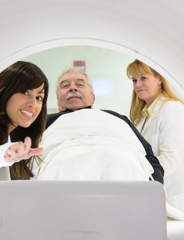
Magnetic Resonance Imaging (MRI): An MRI uses a large magnet, radio waves and a computer to produce pictures of organs and structures in the body and can often show problems not seen by other imaging methods.
Nuclear Medicine: During a nuclear medicine test, a small amount of radioactive substance is injected into the bloodstream, inhaled or swallowed. The substance releases energy and cameras outside the body detect the energy. The cameras and a computer create detailed images of the inside of the body to help identify medical conditions in the earliest stages. Nuclear medicine can also be used to treat cancer and other medical conditions.
Diagnostic Ultrasound: An ultrasound uses high frequency sound waves to produce images of the structures inside the body. It is also known as sonography. Unlike other imaging techniques, ultrasound does not use radiation, so it can safely examine a baby in a pregnant woman, it can also be used:
- Assessment of the thyroid gland in your neck
- Evaluation of the heart and diagnosis of cardiac problems
- Revealing the presence of infection in a specific area of the body
- Determination of abnormal structures/ growth in a particular area Example: Cysts, abnormal growth, tumor etc.
- Revealing abnormalities in the reproductive organs
Bone Sensitometry: This specialized X-ray helps diagnose osteoporosis or determine fracture risk.
Swallow Test: A barium swallow test (cine esophagram, swallowing study, esophagography, modified barium swallow study, video fluoroscopy swallow study) is a special type of imaging test that uses barium and X-rays to create images of your upper gastrointestinal (GI) tract. Your upper GI tract includes the back of your mouth and throat (pharynx) and your esophagus.
Fluoroscopy: Fluoroscopy is a study of moving body structures--similar to an X-ray "movie." A continuous X-ray beam is passed through the body part being examined. The beam is transmitted to a TV-like monitor so that the body part and its motion can be seen in detail. Fluoroscopy, as an imaging tool, enables physicians to look at many body systems, including the skeletal, digestive, urinary, respiratory, and reproductive systems.
Bone Scan: A bone scan is a type of nuclear radiology procedure. A tiny amount of a radioactive substance is used during the procedure to assist in the examination of the bones. A bone scan is done to identify areas of physical and chemical changes in bone and is also used to follow the progress of treatment of certain conditions.
Cardiac Catheterization: Often called cardiac cath, the cardiologist inserts a very small, flexible, hollow tube (called a catheter) into a blood vessel in the groin, arm, or neck and threads through the blood vessel into the aorta and into the heart. Contrast dye may be injected to check blood flow through them. (The coronary arteries are the vessels that carry blood to the heart muscle.) This is called coronary angiography.
Once the catheter is in place, other procedures can be done during or after a cardiac cath such as:
- Angioplasty. In this procedure, your doctor can inflate a tiny balloon at the tip of the catheter. This presses any plaque buildup against the artery wall and improves blood flow through the artery.
- Stent placement. In this procedure, your doctor expands a tiny metal mesh coil or tube at the end of the catheter inside an artery to keep it open.
- Intravascular ultrasound (IVUS). This test uses a computer and a transducer to send out ultrasonic sound waves to create images of the blood vessels. By using IVUS, the doctor can see and measure the inside of the blood vessels.
HeartView Scan (Calcium Scoring)
If you have a family history of heart disease or other factors that put you at risk, you should schedule a HeartView Scan (calcium scoring screening). This 10-minute, non-invasive scan on our state-of-the-art CT measures the amount of plaque in the coronary arteries. Plaque is the substance that builds within the walls of the arteries and can cause a heart attack if the arteries become blocked. Early detection is the key to treating heart disease.
Call (903) 798-7955 to schedule a screening. No physician referral is needed.
Cash price
$75
Advanced Imaging Center
5508 Summerhill Road, Texarkana, Texas 75503
Karen Wacha, Director
(903) 794-9729
[email protected]
Centrally located on Summerhill Road, Wadley Advanced Imaging is convenient and accessible for patients. The staff is committed to providing excellent customer service with next day appointments available. Technology available at this location include:
Philips Achieva HD 3.0T MRI - is a powerful and patient friendly compact whole body MRI system with a wide and open short bore design for comfortable and efficient patient imaging.
Open Bore MRI – the opening of the MRI scanner is 20% larger than traditional MRI systems which is a benefit for claustrophobic patients but offers the same quality images. This is the only Open Bore in the Texarkana area.
Fonar Upright Open MRI - The UPRIGHT® MRI is the only MRI scanner than can scan patients in any position, including sitting, standing, bending or lying down. This unmatched feature is made possible by the scanner´s unique magnet configuration.
CT scanner
Ultrasound
X-Ray
Lung Scan
Lung cancer screening using low dose computed tomography (LDCT) screens patients at high-risk for lung cancer prior to becoming symptomatic. This potential life-saving screening may be right for you if you are a current smoker or have quit smoking within the past 15 years.
Patient Criteria
- Individuals between the ages of 55-77
- History of at least 30 pack years of smoking *(Pack Years is calculated by using Packs per day [20 cigarettes per pack] x number of years smoked - must be at least 30 years)
- A current smoker or former smoker who has quit within the past 15 years
- Patient has not had a chest CT within past 12 months
- Patients who do not have symptoms of lung cancer
For the initial LDCT lung cancer screening, a Medicare beneficiary must have a lung cancer screening counseling and shared decision making visit with a physician or nurse practitioner in order to get the written order for the LDCT.
CT Scan: Frequently Asked Questions
What is a CT?
Computerized tomography (CT) scan uses special X-ray technology to make detailed pictures from different angles of structures in your body. During the test, you will lie on a table that is attached to the CT scanner, which is a large doughnut-shaped machine. Each rotation of the scanner provides a picture of a thin slice of the organ or area.
Should I get a CT scan to screen for lung cancer?
Early diagnosis could save your life. It is important that you speak with your physician to discuss your health history and understand the benefits and risks.
How do I read the results of a CT Scan?
Through the Wadley Lung Scan program, you will receive a written notification on your findings and if follow-up is needed.
A “suspicious” result means the scan shows something abnormal. This could mean lung cancer or it could mean some other condition. You may need to have additional procedures to have a definitive diagnosis.
A “negative” result means that there were no abnormal findings on the CT scan. It does not mean you absolutely do not have lung cancer or that you won’t get lung cancer. Your doctor should discuss when and if you should be tested again.
Does insurance cover CT Scans?
Medicare covers this annual preventive screening if you meet the criteria of being in a high-risk category. If you have commercial insurance, you would need to check with your carrier. There is a cash price if not covered under your insurance of $200.00
For more information to see if you qualify, call 844-490-LUNG (5864)


